
Marc Willinger (ScopeM)
Modern analytical transmission electron microscopes are capable of delivering picometer resolved information about the geometric arrangement of atoms. It is possible to simultaneously obtain quantitative information about the elemental composition of a material and even to measure the local electronic structure or electric and magnetic fields. One of the side-effects of using strongly interacting electrons for the imaging process is the requirement of a good vacuum near the sample and throughout the optical system. Obtaining detailed information about the state of an isolated material in vacuum is not sufficient if we are interested in processes such as material growth- and decomposition, corrosion or (electro)catalysis. With the availability of MEMS- (micro electro mechanic systems) technology based TEM holders for in situ experiments, it is now possible to study the response of a material to a physical or chemical stimuli and study gas-phase, temperature and electrochemically induced processes. Since atomic motions can be fast and the temporal resolution of conventional microscopes is limited and furthermore, processes are often related to collective dynamics of many atomic species, a combination of high-resolution imaging with context embedded observation at lower magnification is required.
In my presentation I will show how the combination of in situ scanning and transmission electron microscopy enables a multi-scale approach for the study of functional materials in their relevant state. Examples range from CVD growth of 2D materials [1] to corrosion processes and catalysed surface reactions (see Figure1) [2]. It will be shown how in situ microscopy reveals the beauty of complex dynamics in systems that are operated far from thermodynamic equilibrium. The field is still relatively new and with the development of more sensitive and faster detectors as well as further improvement of tools for in situ electron microscopy, we are looking towards an exciting expansion of our possibilities to study complex processes related to atomistic dynamics. 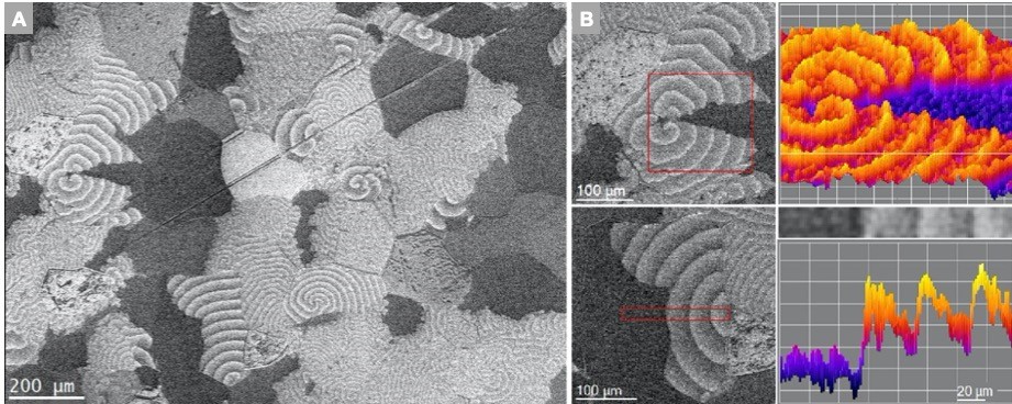 Figure 1: A shows an overview image recorded during NO2 hydrogenation on Pt at T = 172 °C; NO2:H2 ≈ 1:10; ptot = 3.6 × 10⁻² Pa. Propagating chemical waves give rise to beautiful dissipative structures. B shows the variation in contrast between subsequent light-off events and 3D plots of the secondary electron intensity.
References:
- J. Wang et al., ACS Nano, 2015, 9, 1506–1519, Z.-J. Wang et al., Nature Communications, 2016, 7:13256, Z.-J. Wang et al., Adv. Mater. Interfaces, 2018, 1800255, M. Huang et al. Nature Nanotechnology, 2020, doi:10.1038/s41565-019-0622-8
- Barroo, Z.-J. Wang, R. Schlögl, M.-G. Willinger, Nature Catalysis, 2019, doi:10.1038/s41929-019-0395-3